Surgical technique for gama nail
•Download as PPTX, PDF•
1 like•496 views

The biomechanical features of the orthopedic implant gamma nail system offers significantly greater strength and stability compared with the DHS.

Recommended
More Related Content
What's hot
What's hot (20)
Similar to Surgical technique for gama nail
Similar to Surgical technique for gama nail (20)
Recently uploaded
Recently uploaded (20)
Surgical technique for gama nail
- 2. TABLE OF CONTENTS • INTRODUCTION • INDICATION • PREOPERATIVE PLANNING • SURGICAL TECHNIQUE
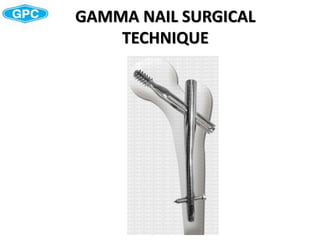
- 3. INTRODUCTION • Gamma Nails come in 3 neck-shaft angles: 120, 125 and 130°. • The anatomical shape of the nail is universal for all indications. • The nail is cannulated for guide-wire-controlled insertion and features a conical tip for optimal alignment with inner part of the cortical bone. • A range of three different neck-shaft angles are available for cephalic screw entry to accommodate variations in femoral neck anatomy. • A single distal Locking Screw is provided to stabilize the nail in the medullary canal and to help prevent rotation in complex fractures. The hole allows for static locking.
- 4. Material: Titanium alloy or Stainless Steel • Nail length: 180mm,200mm,220mm • Nail diameter: proximal 15.5mm, distal: 11.0mm • Proximal Nail angle range: 120°, 125°, 130° • M-L bend for valgus curvature: 4 degrees • End Caps in lengths of 0mm • Distal hole for 6.255mm screws INTRODUCTION
- 5. INTRODUCTION Cephalic Screw and Set Screw Function The Cephalic Screw are designed to transfer the load of the femoral head into the nail shaft by bridging the fracture line to potenitally allow more efficient and more secure fracture healing. The load carrying thread design of the Gamma cephalic screw allows for large surface contact to the cancellous bone. This provides high resistance against cut out. The Set Screw is designed to fit into one of the four grooves of the cephalic screw shaft. This helps prevent both rotation and medial migration of the cephalic screw. The nail allows sliding of the cephalic screw to the lateral side for dynamic bone compression at the fracture site to help enhance fracture healing.
- 6. Technical Specifications • Cephalic Screw diameter: 12.5mm • Cephalic Screw lengths: 70−130mm in 5mm increments •Cephalic Screw is designed for high load absorption and easy insertion • Self retaining Set Screw protects the cephalic Screw against rotation and simultaneously allows for lateral cephalic screw sliding. INTRODUCTION
- 7. INTRODUCTION Distal Locking Screws The distal Locking Screw has a short self-tapping tip which facilitates a faster and easier start as well as easy screw insertion. It helps to promote excellent surface to bone contact Technical Specifications • Distal Locking Screw Diameter: 6.25mm. • Distal Locking Screw lengths ranging from 40−100mm, in 5mm increments. • Fully threaded screw design. • Self-tapping screw tip with optimized short cutting flutes. • Optimized diameter under the head helps to prevent micro
- 8. INTRODUCTION Strength and Stability The biomechanical features of the orthopedic implant gamma nail system offers significantly greater strength and stability compared with the DHS. The Biomechanical Advantage Since the load-bearing axis of the Gamma Nail is closer to the hip joint fulcrum, the effective lever arm on the implant and femur is significantly shorter than with an extramedullary plate. The resultant force is transmitted directly down the femur using a nail system. If DHS is used, the femur shaft may be weakened through a high number of screws. The Gamma Nail increases both the strength and reliability of the biomechanical repair. Rehabilitation Benefits Allows early weight-bearing even in patients with complex or unstable proximal fractures . Early mobilization and a less traumatic operative technique help to increase the chance for more efficient recovery and reliable bone union.
- 9. SURGICAL TECHNIQUE Indications • Intertrochanteric fractures • Nonunion and malunion Contraindications • Medial neck fractures • Sub-trochanteric fractures.
- 10. SURGICAL TECHNIQUE Pre-operative Planning The Gamma Nail with a 125° nail angle may be used in the majority of patients. The 120° nail may be needed in patients with coxa vara, and the 130° nail for coxa valga. The femoral neck angle, (i. e. the angle between the femoral shaft mid-axis and the femoral neck mid-axis) could be measured using a goniometer, to select the apropriate angle of implant to be used.
- 11. SURGICAL TECHNIQUE Patient Positioning The patient is placed in a supine position on the fracture table and closed reduction of the fracture is recommended. Reduction should be achieved as anatomically as possible. If this is not achievable in a closed procedure, open reduction may be necessary. Traction is applied to the fracture to keep the leg straight. The unaffected leg is abducted as far as possible to make room for the image intensifier. While maintaining traction, the leg is internally rotated 10–15 degrees to complete fracture reduction; the patella should have an either horizontally or slightly inward position .
- 12. SURGICAL TECHNIQUE Position the image intensifier so that anterior- posterior and mediolateral views of the trochanteric region of the affected femur can be easily obtained. This position is best achieved if the image intensifier is positioned so that the axis of rotation of the intensifier is centered on the femoral neck of the affected femur. It is important to ensure that a view of both the distal and proximal ends of the nail can be obtained during the procedure without obstruction by the traction table. Patient Positioning
- 13. SURGICAL TECHNIQUE Incision The tip of the greater trochanter may be located by palpation and a horizontal skin incision of approximately 2−3cm is made from the greater trochanter in the direction of the iliac crest. A small incision is deepened through the fascia lata, splitting the abductor muscle approximately 1−2cm immediately above the tip of the greater trochanter, thus exposing its tip. A selfretaining retractor or tissue protection sleeve is put in place.
- 14. SURGICAL TECHNIQUE Entry Point The correct entry point is located at the junction of the anterior third and posterior two-thirds of the tip of the greater trochanter and on the tip itself. The medullary canal has to be opened under image intensification. The use of the curved Awl (CPC622) is recommended.
- 15. SURGICAL TECHNIQUE Preparation of the Medullary Canal Instruments CPC620 Guide Wire Olive FRS68 Flexible reaming shaft RH09 Reaming heads 9, 10, 11, 12, 13 mm
- 16. SURGICAL TECHNIQUE Preparation of the Medullary Canal Guide Wire olive(CPC620) is recommended as a reamer guide. Pass the guide wire into the shaft of the femur using the T Handle. Rotating the Guide Wire during insertion makes it easier to achieve the desired position in the middle of the medullary canal. Flexible reamers are used to ream the shaft of the femur in stages starting from 9mm diameter and increasing in 1mm increments . The canal should be reamed at least 2mm larger than the distal diameter of the nail, 13mm for the 11mmGamma Nail. When reaming is performed, the entire femoral canal should be over-reamed down through the isthmus in order to avoid stress riser in the bone.
- 17. SURGICAL TECHNIQUE In order to accommodate the proximal part of the Gamma Nail, the subtrochanteric region must be opened up to 15.5mm . This can be done by reaming with the reamer. Preparation of the Medullary Canal
- 18. SURGICAL TECHNIQUE Assembly of Targeting Jig Instruments CPC629 Holding jig CPC632 Insertion bolt CPC633 Driver for gamma nail CPC635 Universal Wrench
- 19. SURGICAL TECHNIQUE Nail Assembly The selected Gamma Nail is now assembled to the holding jig(CPC629) as shown in Figure. Ensure that the locating pegs fit into the corresponding notches of the proximal part of the nail. Fully tighten the insertion bolt(CPC632)with the driver for gamma nail and universal wrench, so that it does not loosen during nail insertion. Before starting surgery the following functions of the holding jig have to be checked: 1. Secure fixation between Nail and jig. 2. Lag Screw Guide Sleeve matches the selected nail angle. 3. Distal locking sleeve matches the hole of the distal hole of gamma nail.
- 20. SURGICAL TECHNIQUE Pass the protection sleeve for proximal locking and guide wire sleeve for gamma nail gently through the hole of the jig. Check correct nail angle using the guide wire. Remove the protection sleeve and guide wire. The protection sleeve for distal locking and drill sleeve for distal locking are passed through the jig until its final position is achieved. Check position with the Drill Sleeve and 5.5mm Drill bit. Nail Assembly Check
- 21. SURGICAL TECHNIQUE Nail Insertion Insert the Gamma Nail by hand. Even if some resistance is felt during nail insertion, never hammer to insert the nail, because these high forces will create stress to both bone and to the nail. It may create micro fractures in the bone or deform the nail, which may lead to a reduced targeting accuracy when drilling.
- 22. SURGICAL TECHNIQUE The final nail depth position is monitored with the image intensifier C-Arm; the projected axis of the cephalic screw may be projected with a ruler on the monitor screen to ensure that the cephalic screw is placed in the optimal position. Proceed until the axis of the cephalic screw hole (visible as a crescent shape on the screen) is aligned with the lower half of the femoral neck . The objective is to position the cephalic screw centrally or slightly inferior in femoral head in the frontal plane. The Lag Screw should be placed in the central position of the femoral head in the lateral view Nail Insertion
- 23. SURGICAL TECHNIQUE Cephalic Screw Insertion Instruments PSL794 Protection sleeve for proximal locking PS921 Guide wire sleeve CPC638 Guide wire for cephalic screw IBS755 Pointer for proximal locking WCS800 Tap for cephalic screw CPC626 Cannulated reamer for cephalic screw WC70 Wrench for cephalic screw
- 24. SURGICAL TECHNIQUE Assemble the protection sleeve for proximal locking(PSL794) with the pointer for proximal locking(IBS755) and pass them through the jig to the level of the skin. Make the skin incision down to the bone .When the tip reaches the bone, replace the pointer to the guide wire sleeve(PS921). For an accurate cephalic Screw length measurement, the outer protection sleeve must be in good contact with the lateral cortex of the femur. Insert guide wire for cephalic screw(CPC638) into neck. The objective is to position the cephalic screw in the centre or below the centre of the femoral head in the antero posterior view and centrally in lateral view provides the best load transfer to the cephalic screw. Cephalic Screw Insertion
- 25. SURGICAL TECHNIQUE Cephalic Screw Insertion The guide wire sleeve is now removed and cannulated reamer(CPC626) is passed over the guide wire through the protection sleeve for proximal locking(PSL794). The drilling process, especially when the tip of the drill comes close to its final position in the femur head, should be controlled under an image intensifier to avoid hip joint penetration.
- 26. SURGICAL TECHNIQUE Cephalic Screw Insertion The chosen cephalic screw is then attached to the wrench for cephalic screw(WC70). In a case where compression is to be applied, a shorter cephalic screw length should be chosen to avoid the end sticking out too far in to the lateral cortex . Ensure that the pins of the wrench are in the slots of the cephalic screw. The cephalic screw assembly is now passed over the guide wire, through the protection sleeve for proximal locking, and threaded up to the end of the pre-drilled hole of the femur head. Check the end position of the cephalic screw on the image intensifier.
- 27. SURGICAL TECHNIQUE Cephalic Screw Fixation The handle of the cephalic screwdriver must be either parallel or perpendicular (90°) to the jig to ensure that the Set Screw fits securely into one of the 4 Grooves on the Lag Screw shaft. If the T-Handle is not perpendicular or parallel to the Target Arm, turn it clockwise until it reaches this position. Never turn the cephalic screw Counter clockwise.
- 28. SURGICAL TECHNIQUE Compression / Apposition If compression or apposition of the fracture gap is required, this can be achieved by gently turning the thumbwheel of the wrench for cephalic screw clockwise against the protection sleeve. In osteoporotic bone care must be taken to prevent cephalic screw pullout in the femoral head. The selected cephalic screw should be shorter depending on the expected amount of compression. Cephalic Screw Fixation
- 29. SURGICAL TECHNIQUE Cephalic Screw Fixation Assemble the Set Screw to the Set Screw driver(IBS747). Insert the Set Screw along the opening of the post of the jig and advance it through the insertion bolt pushing the Set Screwdriver. Push the Set Screw Driver down until you are sure, that the Set Screw engages the corresponding thread in the nail. You may feel a slight resistance while pushing down the assembly. Turn the Screwdriver handle clockwise under continuous pressure. Keep on turning the Set Screw until you feel contact in one of the grooves of the cephalic screw.
- 30. SURGICAL TECHNIQUE Cephalic Screw Fixation To verify the correct position of the Set Screw, try to turn the wrench for cephalic screw gently clockwise and counterclockwise. If it is not possible to turn the wrench for cephalic screw, the Set Screw is engaged in one of the grooves. If the wrench for cephalic screw still moves, recorrect the handle position and tighten the Set Screw again until it engages in one of the four grooves. After slightly tightening the Set Screw it should then be unscrewed by one quarter (¼) of a turn, until a small play can be felt at the Lag Screwdriver. This ensures a free sliding of the Lag Screw. Make sure that the Set Screw is still engaged in the groove by checking that it is still not possible to turn the cephalic screw with the wrench for cephalic screw.
- 31. SURGICAL TECHNIQUE Instruments PSL793 Protection Sleeve for distal Locking IBS715.40 Drill sleeve for Distal Locking IBS738 Drill Bit for distal locking IBS747 Screw driver for distal locking Distal Locking
- 32. SURGICAL TECHNIQUE Distal Locking • Disconnect the wrench for cephalic screw loosening the end thumbwheel, remove the wrench, cephalic screw protection sleeve and guide wire. Important points to remember before distal locking procedure: • Ensure that the insertion bolt is still fully tightened • Avoid soft tissue pressure on the distal locking sleeve assembly. Adequate skin incision is important. • Check that the distal locking sleeve assembly (with the trocar removed) is in contact with lateral cortex of the Femur .Confirm final locking screw placement with A/P and Lateral fluroscopy. • Do not apply force to the Targeting jig. • Start the power tool before having bone contact with the drill. • Use sharp drills only.
- 33. SURGICAL TECHNIQUE • Assemble the protection sleeve for distal locking(PSL793),drill sleeve for distal locking(IBS738), and advance it through the hole of the target arm down to skin. • A small incision is started at the tip of the trocar, and is extended down to the lateral cortex. • Insert the protection sleeve(PSL793) and drill sleeve for distal locking(IBS715.40) till the lateral cortex of femur. Distal Locking
- 34. SURGICAL TECHNIQUE • Use drill for distal locking 5.5mmx120mm(IBS738) to drill through first and second cortex. This can be monitored by image intensifier. • Use depth guage to measure length of the distal locking screw after removing the drill sleeve. • Insert the 6.25mm distal locking screw through the drill sleeve for distal locking with the help of screwdriver. Advance the screw head until it is in direct contact with the cortex. Distal Locking
- 35. SURGICAL TECHNIQUE Insert End Cap • It is recommended to use an end cap to close the proximal part of the nail to prevent bone in growth. • Remove the insertion bolt using the driver for gamma nail and universal wrench. • Load the End Cap (0mm) to one of the Screwdrivers and pass the assembly through the top of the jig down into the nail. • Turn the handle clockwise until it stops mechanically. Remove the • Screwdriver and remove the jig in the cranial direction. • Alternatively, the End Cap could be inserted free hand after removal of the jig.