
How to split an axon
Our nervous system is an incredibly complex labyrinth in which single cells are wired up to many others. Building this system requires that the wire-like extension of each nerve cell – called an axon – branches multiple times as it grows. So far little has been known about the process by which this happens. Now Hannes Schmidt and other members of Fritz Rathjen’s group at the MDC have discovered a set of signals within the neuron that are crucial to one type of axon branching. Their work appears in the October 22, 2007, issue of the Journal of Cell Biology.
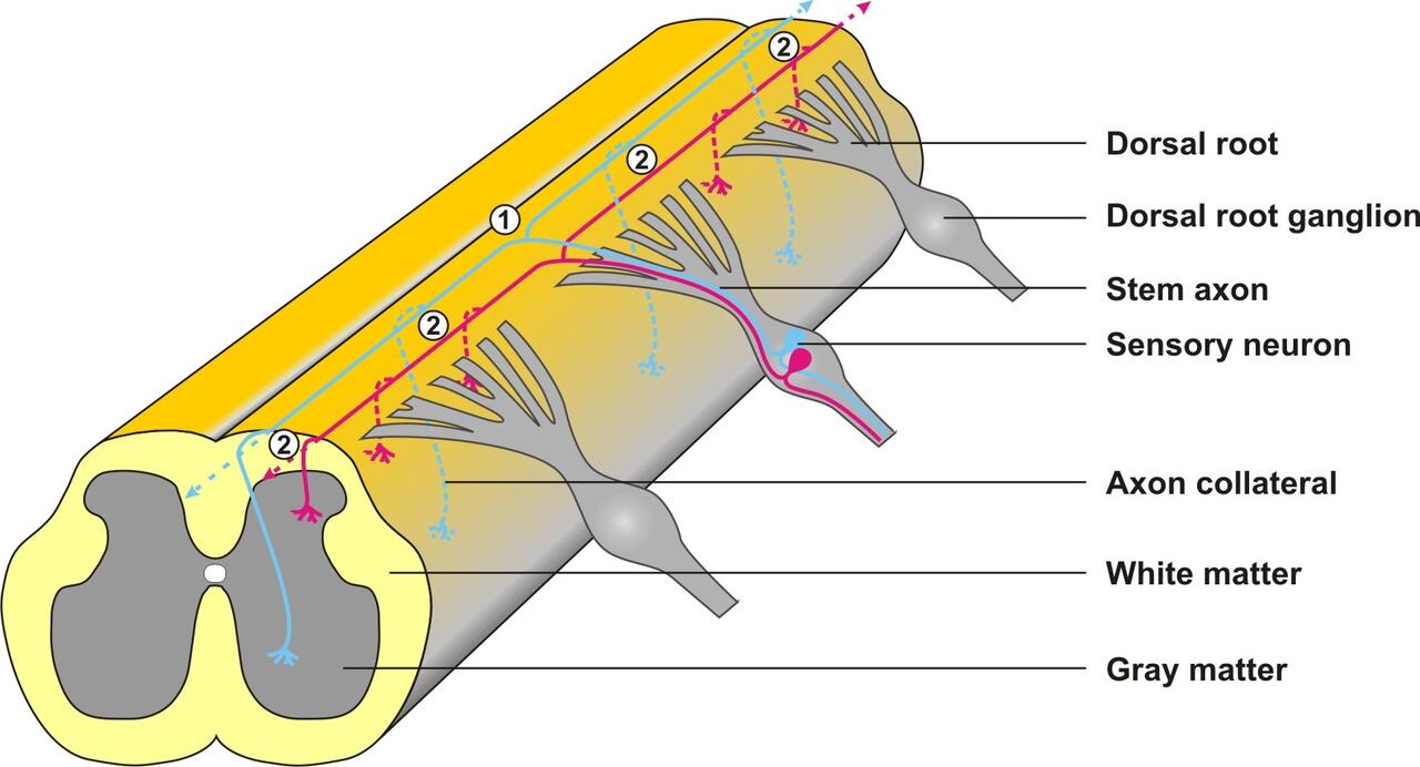
Schematic representation of a sensory neuron. When the axon of the sensory neuron grows into the gray matter of the spinal cord, two types of branching can be observed: At the dorsal root entry zone the axon shaft divides into two branches (1), which continue to grow on the surface of the spinal cord in opposite directions. Out of these branches collaterals then sprout in several places (2) thus enabling the transmission of a signal to several target cells. (Drawing: Hannes Schmidt / Copyright: MDC)
Sensory neurons which sense pain and detect changes in temperature and pressure have to be connected to relay neurons in the spinal cord that transmit the information to the brain (figure 1). As an embryo develops, an axon grows from each sensory nerve cell and finds its way to the cord. When the tip reaches a region called the dorsal root entry zone (DREZ), it splits into a T-shaped fork. The two new tips head off in opposite directions at the border of the spinal cord. Later, branch-like collaterals sprout from these stems; they grow into the gray matter of the cord and connect to relay neurons.
One finding from the new study is that different molecules control the creation of collaterals and forks. While investigating a strain of mouse lacking a protein called Npr2, Schmidt and his colleagues discovered that the animal’s sensory neurons did not make the T-shaped branches. Instead, when the tips of the axons reached the DREZ, they headed one way or the other (figure 2). They were still able to sprout the collaterals, but overall they made fewer connections to other neurons in the spinal cord. This could be observed by making electrophysiological measurements of stimulation within the nervous system.
Why does the loss of Npr2 change the branching of axons? Schmidt and his colleagues have discovered that the signal to make a fork is passed from Npr2 via other cellular information molecules called cGMP and cGKI. In normal mice Npr2 picks up a cue that activates the production of cGMP within the cell. The rise of cGMP levels switches on the cGKI protein, which goes on to activate other molecules. If any of these molecules are absent, T-forking doesn’t take place. As a result, some sensory information is probably lost before it can be transmitted to the brain.
- Russ Hodge


