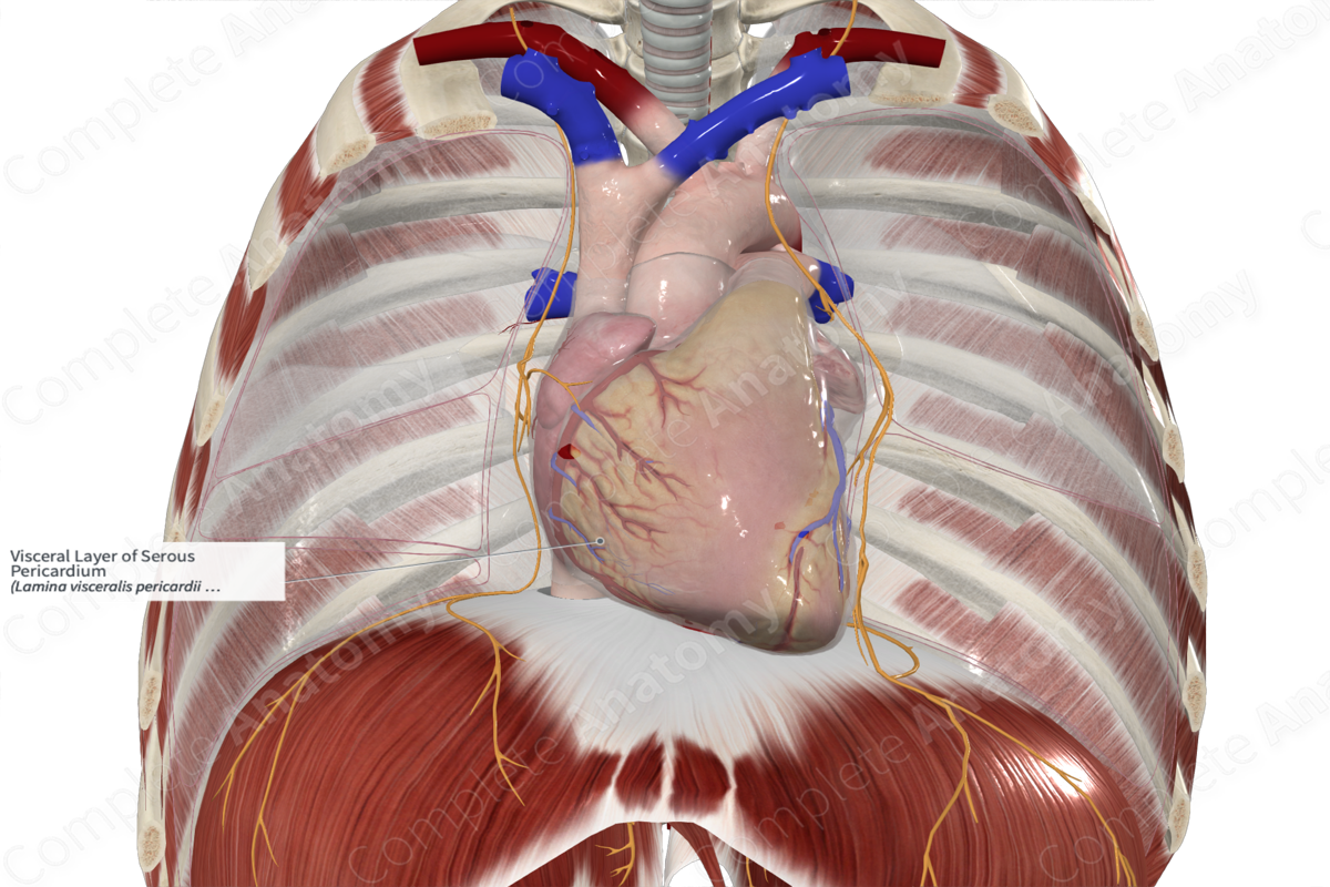
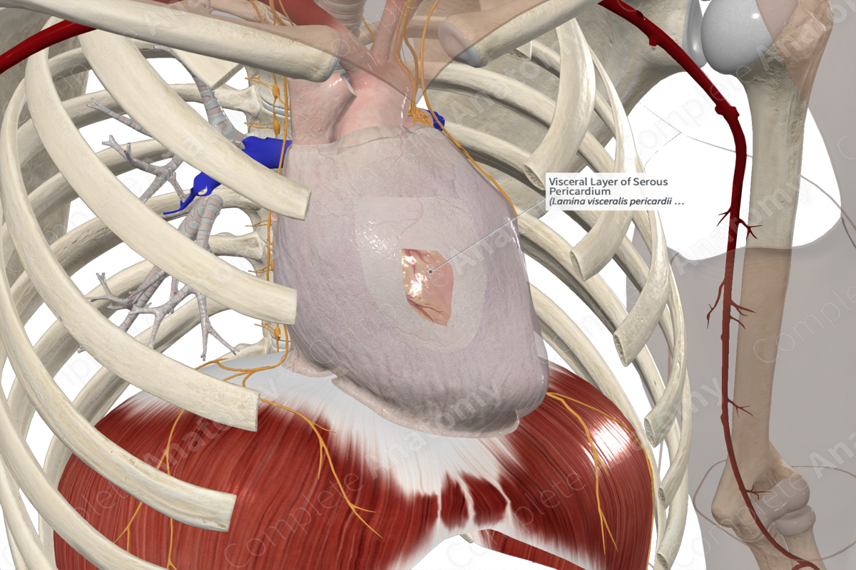
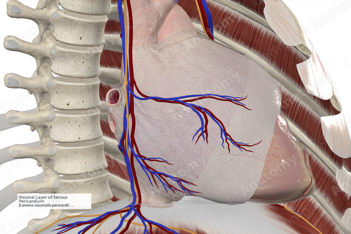
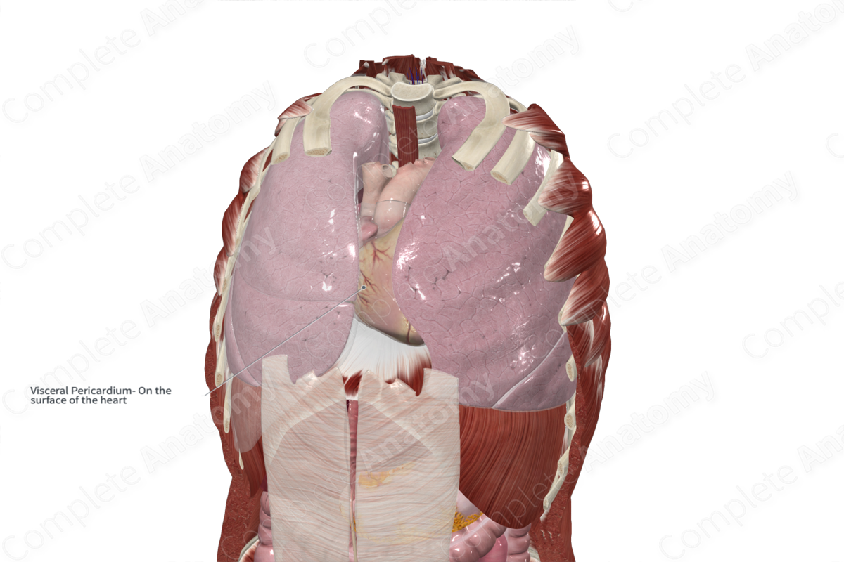
Visceral Layer of Serous Pericardium
Lamina visceralis pericardii serosi
Read moreMorphology/Structure
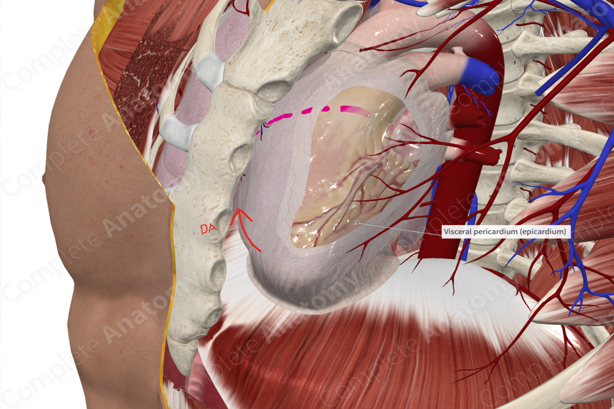
The inner lining of the pericardium consists of two serous layers, parietal and visceral. The visceral layer is the innermost layer and adheres to the cardiac tissue as the epicardium. The parietal layer is attached to the internal lining of the fibrous pericardium. The visceral layer of serous pericardium is innervated by sympathetic (aortic and cardiac plexuses) and parasympathetic fibers from the vagus nerve (CN X); thus, the visceral layer is insensitive to pain.
The serous layers consist of a single mesothelial layer of ciliated cells sitting on a thin connective tissue abundant in elastic fibers and adipocytes. The pericardial sac is formed between the parietal and visceral layers of the serous pericardium.
Key Features/Anatomical Relations
The parietal and visceral layers are continuous around the roots of the great vessels and form two recesses, the oblique and transverse sinuses. The oblique sinus lies behind the base of the heart between the pulmonary veins. Although rarely necessary, the approach to the oblique sinus is to slide your hand behind the heart from the apex so that the bulk of the left ventricle sits in the palm of your hand. Your fingers will be able to slide up until they reach a U-shaped cul-de-sac of pericardium which indicates the posterior aspect of the base of the heart (left atrium).
The transverse sinus is formed by posterior and superior serosal pericardial reflections. It’s posterior to the pulmonary trunk and ascending aorta, superior to the left atrium, and anterior to the superior vena cava. The transverse sinus is an important landmark in cardiac surgery. A finger can be passed through the transverse sinus to insert a surgical clamp for ligation of the arterial supply and insertion of a coronary bypass machine. To find the transverse sinus you may pass a finger posterior to the aorta and pulmonary trunk so that they sit in front of your finger while keeping the superior vena cava behind your finger.
Function
The visceral layer of the serous pericardium aids in the production of serous fluid secreted into the pericardial sac (approximately 20 ml), thus permits friction-free movement of the heart within the thoracic cavity.
List of Clinical Correlates
- Cardiac tamponade
- Pericardial effusion
- Pericarditis
- Pericardial rub
Learn more about this topic from other Elsevier products
Epicardium

epicardium is the connective tissue covering of the heart and corresponds to the tunica adventitia of vessels.
Epicardium: What Is It, Functions, and More

The epicardium refers to the outermost protective layer of the heart
