
16.4 The Cytoskeleton
During cell division, the microtubules of the cytoskeleton are remodeled to form the mitotic spindle. The mitotic spindle, or spindle apparatus , attaches to kinetochores and pulls the sister chromatids away from one another. The mitotic spindle is composed of microtubules, which are part of the cytoskeleton.
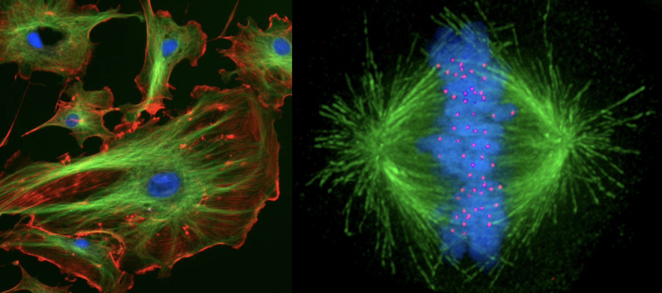
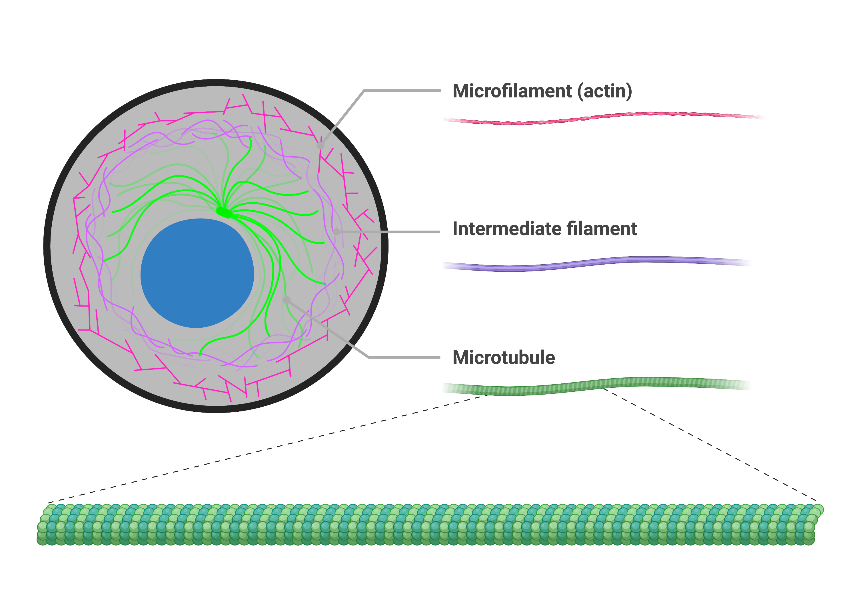
The cytoskeleton is a network of protein fibers that help maintain the cell’s shape, secure some organelles in specific positions, allow cytoplasm and vesicles to move within the cell, and enable cells within multicellular organisms to move. Collectively, scientists call this network of protein fibers the cytoskeleton. There are three types of fibers within the cytoskeleton: microfilaments, intermediate filaments, and microtubules.
Microfilaments
Of the three types of protein fibers in the cytoskeleton, microfilaments are the narrowest. They function in cellular movement, have a diameter of about 7 nm, and are comprised of two globular protein intertwined strands, which we call actin. For this reason, we also call microfilaments actin filaments.

ATP powers actin to assemble its filamentous form, which serves as a track for the movement of a motor protein we call myosin. This enables actin to engage in cellular events requiring motion, such as cell division in eukaryotic cells and cytoplasmic streaming, which is the cell cytoplasm’s circular movement in plant cells. Actin and myosin are plentiful in muscle cells. When your actin and myosin filaments slide past each other, your muscles contract.
Microfilaments also provide some rigidity and shape to the cell. They can depolymerize (disassemble) and reform quickly, thus enabling a cell to change its shape and move. White blood cells (your body’s infection-fighting cells) make good use of this ability. They can move to an infection site and phagocytize the pathogen.
Intermediate Filaments
Several strands of fibrous proteins that are wound together comprise intermediate filaments. Cytoskeleton elements get their name from the fact that their diameter, 8 to 10 nm, is between those of microfilaments and microtubules.
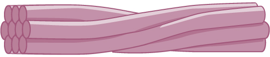
The intermediate filaments are the most diverse group of cytoskeletal elements. Several fibrous protein types are in the intermediate filaments. You are probably most familiar with keratin, the fibrous protein that strengthens your hair, nails, and the skin’s epidermis.
Microtubules
As their name implies, microtubules are small hollow tubes. Polymerized dimers of α-tubulin and β-tubulin, two globular proteins, comprise the microtubule’s walls. With a diameter of about 25 nm, microtubules are cytoskeletons’ widest components. They help the cell resist compression, provide a track along which vesicles move through the cell, and pull replicated chromosomes to opposite ends of a dividing cell. Like microfilaments, microtubules can disassemble and reform quickly.
Microtubules are also the structural elements of flagella, cilia, and centrioles (the latter are the centrosome’s two perpendicular bodies). In animal cells, the centrosome is the microtubule-organizing center. In eukaryotic cells, flagella and cilia are quite different structurally from their counterparts in prokaryotes, as we discuss below.
Flagella and Cilia
The flagella (singular = flagellum) are long, hair-like structures that extend from the plasma membrane and enable an entire cell to move (for example, sperm, Euglena, and some prokaryotes). When present, the cell has just one flagellum or a few flagella. However, when cilia (singular = cilium) are present, many of them extend along the plasma membrane’s entire surface. They are short, hair-like structures that move entire cells (such as paramecia) or substances along the cell’s outer surface (for example, the cilia of cells lining the Fallopian tubes that move the ovum toward the uterus, or cilia lining the cells of the respiratory tract that trap particulate matter and move it toward your nostrils.)
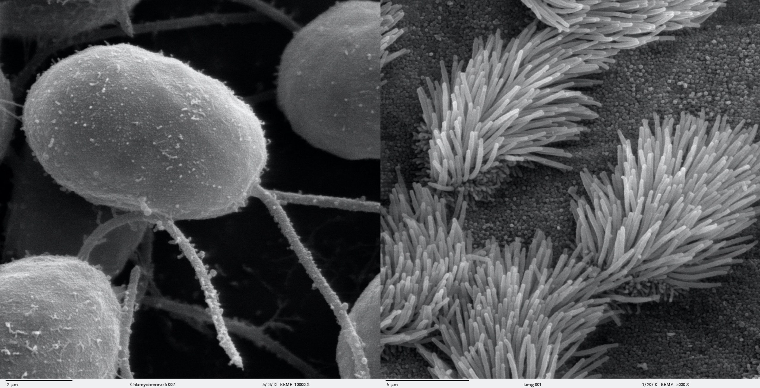
Despite their differences in length and number, flagella and cilia share a common structural arrangement of microtubules called a “9 + 2 array.” This is an appropriate name because a single flagellum or cilium is made of a ring of nine microtubule doublets, surrounding a single microtubule doublet in the center.
apparatus composed of microtubules that orchestrates the movement of chromosomes during mitosis
protein structure associated with the centromere of each sister chromatid that attracts and binds spindle microtubules during prometaphase
protein fiber network that collectively maintains the cell's shape, secures some organelles in specific positions, allows cytoplasm and vesicles to move within the cell, enables unicellular organisms to move independently, and allows some cells to move within a multicellular organism
the cytoskeleton system's narrowest element; it provides rigidity and shape to the cell and enables cellular movements; composed of many actin subunits
cytoskeletal component, comprised of several fibrous protein intertwined strands, that bears tension, supports cell-cell junctions, and anchors cells to extracellular structures
polymers of tubulin proteins that form long fibers; part of the cytoskeleton. Helps the cell resist compression, provides a track along which vesicles move through the cell, pulls replicated chromosomes to opposite ends of a dividing cell, and is the structural element of centrioles, flagella, and cilia
long, hair-like structure that extends from the plasma membrane and moves the cell
short, hair-like structure that extends from the plasma membrane in large numbers and functions to move an entire cell or move substances along the cell's outer surface
