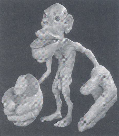
There are so many perspectives from which we can see ourselves. Have you seen yourself from this one? This dysmorphic dude is a representation of the body found within our brains. Here’s the map by which he was made.
- The somatosensory and motor cortices; image borrowed from this post by Antranik.
Nerves from all parts of the body are bundled and pass into the brain, where their presence is registered on the surface of these cortices. Generally speaking, the proportions of the homunculus reflect how much surface area of the cortex is devoted to the given body part. In turn, the cortical area is proportional to the density of innervation at a given place in the body. The bigger the body part, the more densely packed with nerves, and thus the greater its representation on the cortices.
There are two versions of the homunculus: one reflects the sensory cortex and the other the motor cortex. The amount of sensory cortex devoted to a given body part is proportional to the density of cutaneous tactile receptors. The human lips and hands loom large. No wonder we’d rather kiss on the lips (or hands) than rub asses or thighs.
Speaking of thighs, notice how much smaller they are than our thumbs. From the viewpoint of the motor cortex, this is hardly surprising, considering how nimble—indeed how handy—thumbs are compared with the relatively dull and oxlike thighs.
The man who discovered this “body within the brain” and produced the first such map was neurosurgeon Wilder Penfield. By probing the surface of the cortices with electrical probes he discovered that patients felt sensations in different parts of the body. (Having your skull opened and the surface of your brain probed sounds pretty awful, but it turns out it’s not in the least painful.) Penfield pioneered the Montreal procedure, an effective way of treating epilepsy by destroying the nerve cells in the brain where the seizures originate. Before destroying the cells, he’d probe around in order to accurately target the responsible parts, and thus he discovered and mapped the sensory and motor cortices.
Penfield tested only men, so until recently there was no distinctively female homunculus. Now some research has finally filled in the relevant missing parts.
The main findings, summarized in a good post by a (female) Aussie professor, are these
1/ The clitoris is in the same place as the penis
2/ Vaginal stimulation activates different areas of the brain to clitoral stimulation
3/ Nipple stimulation not only activates the chest on the homunculus it also activates the genitals.
Speaking of genitals, they also loom large, for reasons too obvious to need mentioning. Yet their mapping was a delicate matter for 1950s morals, the world in which Penfield operated. (He published his work in 1951.) In a bow to modesty, some versions have the homunculus—Latin for “little man”—in a loincloth, even a diaper.
One final interesting factoid for fellow fans of science-fiction writer Philip K. Dick, author of Do Androids Dream of Electric Sheep? The characters in that story use a household device called a Penfield Mood Organ to dial up emotions on demand.
Which is really the obvious next step, once you’ve got a map of the territory.


Pingback: Neuroplasticity and Exercising – PAINLESS THOUGHTS