Laboratory Studies
Diagnosis of axillary hyperhidrosis is essentially from patient history (see Clinical) and from visible signs of excessive sweating. No useful formal staging or severity scale exists, but the terms mild, moderate, and severe are used in some clinical descriptions. Hund et al have proposed a quantitative definition of axillary hyperhidrosis, but the requirement of a gravimetric assay makes this approach practical only in the research setting.
Laboratory testing may play a vital role in excluding secondary hyperhidrosis from causes such as hyperthyroidism, pheochromocytoma, carcinoid or other malignancy, tuberculosis, or adrenal pathology, especially in patients with asymmetrical, late, or atypical onset of symptoms.
Other Tests
Heckmann et al have described a gravimetric method for quantitating sweat production in which filter paper is weighed dry on a high-precision laboratory scale, then placed in contact with a hyperhidrotic area of patient skin for 60 seconds, then weighed again. They found the rate of sweat production in hyperhidrotic areas to be near 200 mg/min. [6]
Minor's iodine starch test has been used for many years to map the areas of axillary hypersecretion. [7]
The axilla is dried thoroughly, painted with an iodine tincture, then air-dried. See the image below.
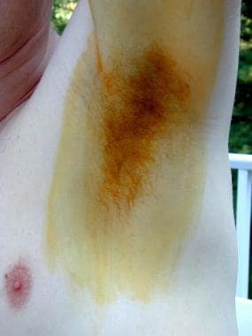 Surgical treatment of axillary hyperhidrosis. Iodine/starch test: Iodine tincture or Betadine applied and air dried. Image courtesy of Richard H S Karpinski, MD.
The dried area is then dusted with cornstarch or potato flour. See the image below.
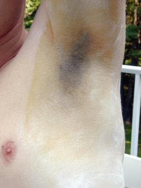 Surgical treatment of axillary hyperhidrosis. Iodine/starch test: Cornstarch powdered onto dried iodine. Image courtesy of Richard H S Karpinski, MD.
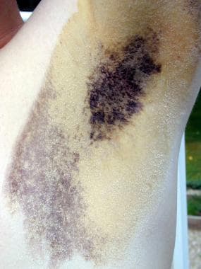
As the patient begins to sweat, moistened starch combines with the iodine to produce a vivid blue color. See the images below.
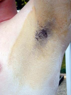 Surgical treatment of axillary hyperhidrosis. Iodine/starch test: As sweating begins, iodine and starch react wherever dampened to produce a blue color. Image courtesy of Richard H S Karpinski, MD.
 Surgical treatment of axillary hyperhidrosis. Iodine/starch test: Further color development (see Image 6). Image courtesy of Richard H S Karpinski, MD.
The hyperhidrotic areas then can be mapped and outlined with an indelible felt-tip marker. See the image below.
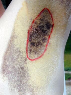 Surgical treatment of axillary hyperhidrosis. Iodine/starch test: An indelible marker outlines the area positive for hyperhidrosis. Image courtesy of Richard H S Karpinski, MD.
A variation of this test uses ninhydrin solution sprayed on an air-dried axilla, relying on color reaction with proteinaceous sweat to produce a visible pattern.
In practice, gravimetric measurement of sweat production is seldom used. Iodine mapping is also of limited usefulness as a prelude to surgery, since the hyperhidrotic area usually corresponds quite closely to the hair-bearing area of the axilla; however, mapping may be extremely helpful in pinpointing an area of recurrence (or failed gland resection) in patients who require reoperation. See the image below.
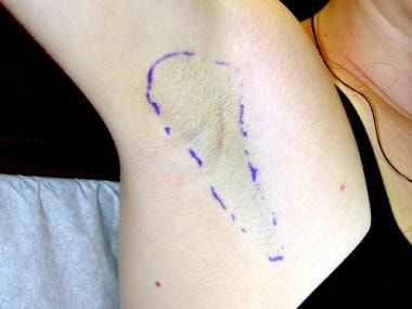 Surgical treatment of axillary hyperhidrosis. Modified Skoog procedure: Outline of the operative site as estimated by the hair pattern (not by mapping). Image courtesy of Richard H S Karpinski, MD.
-
Surgical treatment of axillary hyperhidrosis. Staggered-cross incision of a classic Skoog procedure. Image courtesy of Richard H S Karpinski, MD.
-
Surgical treatment of axillary hyperhidrosis. Karpinski modification of the classic Skoog procedure incision. Image courtesy of Richard H S Karpinski, MD.
-
Surgical treatment of axillary hyperhidrosis. Iodine/starch test: Iodine tincture or Betadine applied and air dried. Image courtesy of Richard H S Karpinski, MD.
-
Surgical treatment of axillary hyperhidrosis. Iodine/starch test: Cornstarch powdered onto dried iodine. Image courtesy of Richard H S Karpinski, MD.
-
Surgical treatment of axillary hyperhidrosis. Iodine/starch test: As sweating begins, iodine and starch react wherever dampened to produce a blue color. Image courtesy of Richard H S Karpinski, MD.
-
Surgical treatment of axillary hyperhidrosis. Iodine/starch test: Further color development (see Image 6). Image courtesy of Richard H S Karpinski, MD.
-
Surgical treatment of axillary hyperhidrosis. Iodine/starch test: An indelible marker outlines the area positive for hyperhidrosis. Image courtesy of Richard H S Karpinski, MD.
-
Surgical treatment of axillary hyperhidrosis. Modified Skoog procedure: Preoperative axilla in a healthy young writer with axillary hyperhidrosis. Image courtesy of Richard H S Karpinski, MD.
-
Surgical treatment of axillary hyperhidrosis. Modified Skoog procedure: Outline of the operative site as estimated by the hair pattern (not by mapping). Image courtesy of Richard H S Karpinski, MD.
-
Surgical treatment of axillary hyperhidrosis. Modified Skoog procedure: Anesthesia is attained by infiltration of local anesthetic. Image courtesy of Richard H S Karpinski, MD.
-
Surgical treatment of axillary hyperhidrosis. Modified Skoog procedure: The transverse incision is made, here exposing the subcutaneous sweat glands. Image courtesy of Richard H S Karpinski, MD.
-
Surgical treatment of axillary hyperhidrosis. Modified Skoog procedure: Through the rather limited incision, dissection is carried to the entire outlined area. The dissection is deep to the glands but superficial to axillary fascia. Image courtesy of Richard H S Karpinski, MD.
-
Surgical treatment of axillary hyperhidrosis. Modified Skoog procedure: In the recess of the incision, the shiny and somewhat striated axillary fascia is visible. In conducting the operation, no part of the dissection should violate this fascia. Image courtesy of Richard H S Karpinski, MD.
-
Surgical treatment of axillary hyperhidrosis. Modified Skoog procedure: Once the flaps are elevated and hemostasis is achieved, the flaps are everted and the layer of the glands snipped off the undersurface of the dermis. Here, the upper portion of the flap has been cleared of sweat glands, while the gland lobules are still visible on the lower portion of the flap. Image courtesy of Richard H S Karpinski, MD.
-
Surgical treatment of axillary hyperhidrosis. The carpet of glandular tissue is seen intact on the left side of the skin flap, while the glands have been resected on the right side of the flap, revealing the underside of the dermis. The axillary fascia is visible as a glistening sheet above the skin flap. Image courtesy of Richard H S Karpinski, MD.
-
Surgical treatment of axillary hyperhidrosis. Modified Skoog procedure: Once the skin flaps are cleared of sweat glands, the dermis should be visible along with the bulbs of many hair follicles. Although not seen clearly in this photo, the fine vessels of the subdermal plexus should be visible under magnification as a network of red vessels. The posture of everting the flap over the surgeon's finger to facilitate gland dissection is demonstrated. Image courtesy of Richard H S Karpinski, MD.
-
Surgical treatment of axillary hyperhidrosis. Modified Skoog procedure: At the completion of gland resection, vascularity of the flaps should show no compromise. Hemostasis should be meticulous. Image courtesy of Richard H S Karpinski, MD.
-
Surgical treatment of axillary hyperhidrosis. Modified Skoog procedure: A drain has been led out through a small incision at the most proximal area dissected. This spot will be the most dependent when the patient is up and about. Image courtesy of Richard H S Karpinski, MD.
-
Surgical treatment of axillary hyperhidrosis. Modified Skoog procedure: The incision has been closed in two layers, and the drain secured with a single suture. The needle end of the drain can be inserted into a Vacutainer red top tube to supply gentle suction once the incision is closed. Image courtesy of Richard H S Karpinski, MD.
-
Surgical treatment of axillary hyperhidrosis. Incision is closed and drain is in place. Image courtesy of Richard H S Karpinski, MD.
-
Surgical treatment of axillary hyperhidrosis. Modified Skoog procedure: The bandage should be padded and should overlie the entire dissected area. The distal portion can be held in place by wrapping the arm, and the proximal portion needs to be attached to the chest so that the dressing acts like a hinge when the arm is abducted. Image courtesy of Richard H S Karpinski, MD.
-
Surgical treatment of axillary hyperhidrosis. Modified Skoog procedure: The completed dressing, in this case using a self-adherent stretchy flexible bandage on top of surgical paper tape and Tegaderm. Image courtesy of Richard H S Karpinski, MD.
-
Surgical treatment of axillary hyperhidrosis. Modified Skoog procedure: Resulting scar at 2 months after surgery. Note the normal texture and appearance of axillary skin and the normal hair pattern. The pink coloration usually is gone at 4-5 months. Image courtesy of Richard H S Karpinski, MD.
-
Surgical treatment of axillary hyperhidrosis. Professor Tord Skoog (1915-1977).