This page provides detailed and updated information about OPG (Panoramic scan).
You can select the topics that interest you from The Topic Menu.
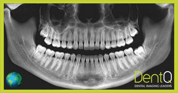
What is an OPG scan?
Orthopantomography (OPG) also known as “Panoramic radiography” is a radiologic technique that provides an overview of the jaws and surrounding structures, in two dimensions. It is one of the most indicated radiographic examinations by dentists as it can give a general overview of entire dental-maxillomandibular structures on a single film. Instead of many advancements in imaging today, panoramic radiography still holds its position intact with great importance.
For patients it is a simple procedure, has relatively low radiation exposure, and is cost-effective. However, image projection is a complex phenomenon as there are multiple superimpositions of the structures in and around the jaws with further distortions due to technical errors in image acquisition. But once a successful interpretation is done it establishes the basis of diagnosis and related treatment.
Why is an OPG test done?
OPG (panoramic scan) involved the usage of x-ray radiation. Therefore, like similar procedures that use x-ray radiation, it required a written medical justification by a doctor. Proper justification for an OPG scan may include the need to investigate various maxillofacial indications and medical conditions, such as:
- Unerupted/Impacted 3rd molars or canines
- Large lesions (tumors, Cysts, etc.)
- Traumatic injuries/ fractures
- Developmental abnormalities
- Tooth development
- Orthodontic treatment
- Implant planning
- Anatomical variations (bifid mandibular canals, retromolar canal, calcified stylohyoid complex) that are rarely noticed but very critical for surgical procedures
OPG (panoramic scan) is also an excellent imaging modality for patients who cannot open their mouth (post-surgical trismus, or trismus due to trauma or pathology).
Is OPG X-RAY painful?
Some radiological procedures require interventional approaches where various small tools, such as catheters, probes, or wires are inserted into the patient’s body during the scan. Even for the intraoral radiographic procedure, x-ray films are placed inside the patient’s mouth. These techniques are sometimes not comfortable for patients and might be painful too.
However, OPG is an extraoral radiographic technique where there is no contact between the patient and the film. Moreover, the time taken by the technique is only 2-3 minutes, which further adds up to the ease of the technique, rather than being painful. The bottom line is that OPG (panoramic scan) procedures are noninvasive, seamless to the patient, and not painful at all.
OPG Procedure
The procedure includes the following steps:
- Patient preparation: the patient is asked to remove any metal objects, jewelry or eyeglasses if wearing.
- Machine preparation: the machine is turned on and the rotating component of the machine, comprising the x-ray source and receptor, are tuned to the start point. Projection parameters are set on the machine based on area of interest, age of patient and reason for scan.
- Patient positioning: patient is asked to stand still with his/her face resting on a small shelf in front. Then they must bite gently on a mouthpiece to keep the head steady during the scan and hold the tongue back while the scan is made. Height of the machine is set to the approximate height of the patient. During the scan, patient positioning is very critical because the quality of the image very much depends on it.
- Image capturing: When the exposure is made the x-ray machine emits x-rays which are directed onto the teeth, bones as well as soft tissues inside the mouth. The structures absorb x-rays based on their composition, the denser ones like teeth and jaws absorb more x-rays and appear clearer on the image whereas the less dense ones like soft tissues and the areas where infections or decay are present absorb fewer X-rays and appear as dark spaces on the image. This helps the radiologist diagnose the disease.
- Data Storage: Entering the patient’s details in a proper database or software for image identification.
- Processing: Verifying that the scan is successful by the radiographer, and adding measurements, observations and recommendations by a radiologist (written in a radiologist report) and sometimes by an artificial intelligence (AI) application as well.
Typically, the entire visit at DentQ and CTdent branches, including admission, preparations, scanning and releasing the patient, takes about 30 minutes altogether for visits that were booked in advance. The scanning itself takes only 2-3 minutes.
OPG Scanners
Basic components of any panoramic machine include the following:
- X-ray tube, which is mounted on one side.
- Detector or receptor on the other side.
The patient’s head is positioned between these two components with the chin and the forehead resting, on either side of the machine also there are handles for support on either side.
- Panoramic machines can have two types of receptors, film based or digital one. The film-based receptor uses a conventional x-ray film that is placed inside the cassette with an intensifying screen, whereas the digital ones are made of photo-stimulable phosphor plate (PSP).
- The receptors can further be divided into two types based on the moving mechanisms of films, one using a sliding flat cassette that holds the film, and another using a rotating cylinder around which the film is wound.
- The machines can be adjusted based on the patient’s height. Some machines have the capability of automated exposure control, where the amount of radiation passing through the patient is calculated during the initial part of the scan and adjustments are made automatically in the latter half of the scan by the machine itself by adjusting the image factors (kVp, mA, and the speed of the imaging movements).
- There can also be an option of adding the cephalometric component in the OPG unit, using which standardized skull views can be obtained with appropriate exposures.
- In cases where multidirectional tomography is involved, the advanced machines that have computer-controlled multimodality can control the direction and speed of movement of the tube head and film.
The OPG machines can capture different views of the TMJ, sinuses, and jaws with this functionality.
OPG devices are created and marketed by various brand, including for example:
- Sirona – top quality, low maintenance, remote serviceability, digital output, price range between INR 1 to 3 lakh
- NewTom and MyRay brands – both manufactured by Cefla, excellent quality and software at mid-range pricing
- Carestream, Vatech – two different brands offering high quality, great ease of use and convenient pricing
- Acteon – a relatively compact and efficient design at convenient pricing
- Additional brands to consider – Kavo, Kerr, 3Shape, Dentium, Fussen and many other
While solely OPG machines are common in some countries, more and more dental clinics and dental imaging centers add Cephalometric arms to the machine, or purchase devices that combine CBCT (3D) and Panoramic (2D) scans in one CBCT device. This trend also means that some of the leading brands, including some those mentioned above, no longer offer standalone OPG machines, but rather combine OPG functionality within CBCT machines.
CBCT machine and OPG
The CBCT and OPG machines are different in their basic composition. OPG is a 2D radiographic technique in which the source of x-rays is a wide beam, whereas CBCT is a 3D radiographic technique in which the source of x-rays is a cone-shaped beam.
The second basic component is the receptor, in CBCT the receptor can produce high-quality cross-sectional images as compared to OPG.
On a routine basis, OPGs are used for dental assessments for the initial stage due to their cost effectiveness, whereas CBCTs give a 3-dimensional view that is very detailed, and the CBCT scan is costly too.
As mentioned above, usually CBCT machines include built-in OPG functionality, and therefore can replace a stand alone OPG machine, and be used both for CBCT (3D) scans and Panoramic (2D) images.
How to read an OPG scan?
OPG scan is a 2D representation of 3D structures. Reading an OPG is very challenging because there are many anatomic structures present in the midface area that superimpose on each other, and there are artifacts too due to several reasons (for example – ray scattering due to metal fillings and crowns) that add to the difficulty of reading the scan.
To read a scan, the starting point is to have a systematic analysis of the image and a thorough understanding of the appearance of normal anatomic structures and their variations. The digital scan is read on a computer screen, and the conventional radiograph should also be viewed on a viewing box for better clarity. The left and right sides of the patient should be identified clearly on the scan image.
One of the most basic and important aspects of reading a scan is proper numbering of the teeth, followed by determining the area of interest and writing a detailed description for that area. While reading a scan, any incidental finding that is present should be also included in the report and stated clearly.
OPG Interpretation
The OPG (Panoramic) scan gives a panoramic image of many structures present in the midface area, along with artifacts and ghost images. Therefore, interpretation of OPG is a critical subject and must be done carefully.
The images obtained on the OPG can be categorized as follows:
- Real or actual images of:
- Hard tissue: teeth, jaws, the styloid process, the hyoid bone, the orbital rim, the nasal septum, and the base of the skull.
- Soft tissues: earlobes, soft palate, dorsum of the tongue, lips, cheeks, and nasolabial fold.
- Air spaces: oropharynx, maxillary sinus.
- Ghost or artifactual images:
- Cervical vertebrae, palate, or metallic objects
In order to avoid mistakes, many dentists consult with dental radiologists and/or AI (Artificial Intelligence) applications in order to properly interpret the OPG scan. At DentQ and CTdent we provide Human+AI radiology reports for OPG scans, as a service, which is often included in the scanning price.
OPG scan cost
OPG is a widely used dental imaging modality. It is readily available in most of the standardized healthcare facilities, as well as in many dental clinics.
The cost of the scan varies from place to place. Local prices vary from facility to facility pending on pricing policy, added value services, competition, regional taxes, demographics, agreements with insurance companies and supply and demand in a specific area.
According to an internal survey we did at DentQ, typical price range in 2022 in various countries is described in the following table:
| Country | OPG scan Low Prices |
OPG Scan Medium Prices |
OPG Scan High Prices |
Remarks |
| USA | $ 60 | $ 100 | $ 150 | OPG scans cost less when insurance covers it |
| Brazil | $ 20 | $ 30 | $ 40 | 50% reduced OPG scan prices via insurance |
| UK | £ 45 | £ 54 | £ 65 | Referral to CBCT more common than to OPG |
| Germany | € 53 | € 67 | € 87 | OPG done in-house, external scans are mostly CBCT |
| Spain | € 20 | € 30 | € 40 | 50% of external scans are OPG, 50% CBCT (3D) |
| France | € 20 | € 20 | € 20 | Covered by insurance |
| Italy | € 30 | € 40 | € 50 | Higher prices in north Italy, lower in the south |
| Portugal | € 10 | € 17 | € 40 | Relatively low prices for Western Europe |
| Greece | € 26 | € 36 | € 45 | OPG usually done externally (not at clinics) |
| Romania | RON 60 | RON 79 | RON 100 | Prices higher in Bucharest, lower elsewhere |
| HK | HK$ 300 | HK$ 350 | HK$ 400 | Many clinics do it in-house |
| Dubai | € 53 | € 78 | € 106 | OPG scans cost less when insurance covers it |
| Israel | NIS 120 | NIS 145 | NIS 175 | Expect lower prices when insurance covers it |
One thing to take into consideration is that some imaging service providers offer discounts on bundled packages, for example on combo deals for OPG scan and Intraoral scan. However, these combo deals are more common for CBCT (3D) scans, rather than for OPG (2D) scans.
At DentQ and CTdent branches around the world, we enjoy our size advantage to reduce costs. As a result, our strategy is to offer more for less: premium service and quality at reasonable (average and below) costs. Therefore, you can expect our official pricing to be around medium prices. In addition, we have agreements with dentists that allow them to give a discount to their patients, and we also distribute coupons and have special offers from time to time.
What is the difference between OPG and CBCT scan?
OPG (Orthopantomogram), and CBCT (Cone Beam Computed Tomography) are two different types of dental imaging techniques that are each used to produce images of the teeth, jaws, and surrounding structures.
An OPG is a 2D imaging procedure that captures a wide, two-dimensional image of the entire mouth in a single scan. It can be used to detect issues with the position of the teeth, their growth and development, the jaw, or the temporomandibular joint (TMJ). It is also helpful in planning and assessing orthodontic treatment.
CBCT, on the other hand, is a 3D imaging technique that uses a cone-shaped x-ray beam to capture a series of cross-sectional images of a specific area of the mouth. The images obtained are extracted from a three-dimensional DICOM file and are more accurate. CBCT can be used to view individual teeth or the roots of the teeth. It is highly recommended for implant planning, as it enables to assess the quality, quantity and density of the bone in the jaw, for making guides for dental implants, and it helps identify the precise root canal location (in 3D) – often crucial for performing a successful implant procedure.
As for the cost – although both procedures take almost the same time for the scanning itself (2-3 minutes) – CBCT requires more experienced radiographer and extra processing work. In addition, CBCT scan often requires a report by a radiologist, and may need a more expensive machine to perform the scan. Therefore, the cost of CBCT scan for 1 jaw is usually 3-fold, and the cost of CBCT scan for 2 jaws usually cost 5-fold, when compared to the cost of OPG scan (which scans the entire mouth).
As for safety – both panoramic and CBCT scans are pretty safe when justified, but OPG requires less radiation dose since it only scans one plane (2D) whereas CBCT scans multiple layers (3D). To counterbalance this, sometimes the findings of OPG make it necessary to do an additional scan, this time 3D CBCT scan, to provide the needed information to the dentist. For example, when the scan is used for orthodontic treatment, the findings of OPG scan may not suffice, when (in 20% of the cases) an impacted tooth is involved. In such cases, the radiation doses include that of the initial OPG scan plus that of following CBCT scan, which means that taking only a CBCT scan alone, to begin with, was actually a safer (and faster) option. Cephalometric scans present a similar dilemma – the default is 2D scan for Ceph exam (OPG), but 3D scans may be needed as they provide more accurate information, as well as clear image of both sides of the head.
To sum up, OPG scans are two-dimensional, less expensive and not so accurate, whereas CBCT scans are three-dimensional, more expensive and way more accurate. A dentist can select the type of scan based on the specific purpose and the availability of the imaging modality. OPG is more readily available in-clinic compared to CBCT, while CBCT is more recommended for Implant planning.
What is the difference between OPG and Dental scan?
An OPG (Orthopantomogram) is a specific type of dental scan that uses x-rays to produce a detailed two-dimensional image of 2D structures in a single scan, including teeth, jaws, TMJ, sinuses, and surrounding structures.
A dental scan is a general term that can refer to any type of dental imaging technique used to produce images of the teeth, jaws, TMJ, sinuses, and surrounding structures. OPG scans, as well as other types of x-ray or imaging techniques such as CBCT (cone-beam computed tomography), MRI (magnetic resonance imaging), or body CT (computed tomography) scans, are examples of various dental scans.
The difference between the other dental scans and OPG scans is the area that can be covered. OPG is a wide, two-dimensional image of the entire mouth in a single scan, whereas other types of dental scans, such as IOPA and bitewings have limitations in terms of area covered (in example – it often covers only 1-3 teeth).
As mentioned in the previous section, CBCT scans, on the other hand, represent a different type of dental scan, used to provide more detailed or specialized images (when compared with OPG images), and of various areas of the mouth (including 1-3 teeth, to two jaws, for example).
Thus, OPG scan is just one type of scan included in the large variety of dental scan solutions, and the specific type of dental scan that is to be used entirely depends on the type of information that is needed, and the specific treatment involved.
Is OPG (Panoramic scan) safe?
During any dental treatment, whenever an x-ray is required and justified, the first thing that worries patients is exposure to radiation. Worries about radiation exposure are even more common when it comes to OPG. But in reality, the exposure to radiation caused by OPG is significantly less than that caused by intraoral radiographs of the full mouth.
The exposure can be further reduced with the use of a lead apron. However, there will be some radiation exposure while taking a panoramic radiograph, but the amount will be minimal.
When compared to other x-ray examinations, the effective dose for OPG test is 3 orders of magnitude lower. For example, chest CT requires 8 mSv, which is more than 1,000-fold effective x-ray dose compared to an OPG scan, which requires only 0.007 mSv.
In addition, dental radiation has always been considered safer than other forms of medical radiation because it is directed to areas of the body that just aren’t radiosensitive. And if we shield the areas of concern—like the thyroid—and make sure that the lenses of the eyes, and are kept out of the x-ray fields, then we are helping the patient to feel safe and protected.
It is also important to take into account that the human body is used to getting bombarded with x-ray radiation from cosmic rays from space and radon gas in the air constantly from the moment we are conceived to the moment we die, so our bodies are used to repairing the damage caused by low amounts of radiation.
The bottom line is that radiation from dental exams, and even more so for OPG exams, has never been scientifically proven to raise the risk of cancer incidence.
OPG scan in pregnancy
During pregnancy, if possible, X-rays should be avoided!
This is especially true and important in the first trimester of the pregnancy, when all the elective treatments that require x-ray exams probably should be postponed. However, if there is an urgent procedure that cannot be delayed until delivery and if it is mandatory to take an x-ray for such treatment, then it can be taken. Further care can be taken by making the patient wear a lead apron during the scan.
As for the second trimester, it is considered to be a safer period during pregnancy, when dental scans can be done. Again, a lead apron during the scan is advised. And, of course, such dental scan exams must comply with local regulation.
Specifically, OPG scan involves a relatively very-low amount of x-ray radiation, and it is pretty common to take OPG exams during the second trimester. Therefore, at DentQ and CTdent we indeed often perform OPG exams to pregnant women during their second trimester.
Who works with OPG
OPG (orthopantomography) is a type of dental imaging technique that is used to produce an entire image of the lower and upper jaws. This technique is used for the diagnosis and treatment planning of a variety of conditions. Dental professionals such as dentists, dental assistants, trained dental nurses and dental radiographers are the ones who apply this technique. The individual who is responsible for using OPG is completely trained in positioning the patient and operating the OPG machine.
The regulation in this regard varies from country to country, so it is important to confirm that whoever is working with the OPG machine is allowed to do so by the authorities. For example – in some countries dental nurses are not allowed to perform dental scans, and in other countries even dentists are not allowed to do it before completing a radiography course, or other types of specialized training and education.
In this respect, it is also important to confirm that whoever is doing the interpretation of the OPG scan results, is qualified by the medical authorities to do so. Again, in some countries, only radiologists are allowed to provide radiology reports to patients, whereas dentists can only use the scans for their own treatment plan, but not to create and provide radiology reports to their patients (unless they took special dental radiology training beforehand).
Where is OPG done?
An OPG, is one of the routinely used dental imaging techniques that produces a wide image of the upper and lower jaws, which can be used for diagnosis in the initial phases. An OPG is usually done in a dental clinic or a diagnostic imaging center. However, the use of OPG for educational purposes in dental schools makes it mandatory to have an OPG machine in dental colleges and in hospital set-ups.
In different countries, OPG machines scans may be very common in-house at the clinic (e.g. – Germany), whereas in other countries OPG scans are almost always done externally (e.g. – Greece), at hospitals, medical imaging centers and dedicated dental imaging centers (like DentQ and CTdent).
In addition, in some countries it is very common that dentists send patients to do a scan with a colleague – a nearby dental clinic that holds an OPG or CBCT device (for example – in HK). This is not always recommended for the dentist, since some of his patients may decide to stay with the clinic they were sent to and do the needed treatment there. Sending patients to independent dental imaging centers (such as DentQ and CTdent branches in many countries) makes more sense for the dentist in this respect, as he will lose less patients to his competitors.
Who should decide where the OPG is actually done?
One of the questions we often face is “Shouldn’t the patients’ decide where to do their external dental scans, including OPG?”. Our answer in this matter is a clear and sounding “No!”. Explanation follows.
Dental Imaging is a professional service designed to provide insights and answers to dentists and medical professionals. Dental imaging serves as “an extended pair of eyes” for the dentist. On the other hand, patients cannot differentiate between high and low quality dental imaging services.
Patients do not choose the dentist-chair, the implant brand, or the bone-graft type for their treatment. And like all these professional components of the dental treatment, dental imaging should be regarded as a professional tool as well. Therefore, OPG service providers (and providers of all other dental scans) should be chosen by medical professionals, based on their professional experience, and not by the patients. Patients’ voices are very important in many respects, but we strongly believe that this is not one of them.
Software readers for OPG
An OPG is a detailed dental image that shows the entire maxillo-mandibular complex. There are many challenges that a dental professional faces in reporting the OPG scan. In order to view and interpret these images correctly, a dental professional should be thoroughly trained to use the specialized software that is used for reading OPG scans.
There are many different software programs that are specifically designed for reading and analyzing OPG images. Some examples of these programs include Panoramic Viewer, 3D Imaging Software, and DICOM Viewer. These programs allow dental professionals to view the images produced by the OPG, to take digital measurements online or manually on printed slides, and to analyze the scans in order to diagnose the dental condition and further treatment planning.
Can OPG be used for Implants?
OPG can be a useful imaging modality for implant planning. If we start from basics, it is one of the best suited dental imaging modalities which can give a complete view of the entire arch, thus the missing teeth and its adjacent teeth can be viewed and compared for the treatment planning. The OPG scan gives a dental surgeon a basic idea about the bone available for the implant placement in terms of height. In addition, vital structures in the proximity such as inferior alveolar nerve, mental foramen, incisive canal, maxillary sinus, nasal fossa and the adjacent teeth can be seen on OPG.
Nevertheless, OPG scan has inherited limitations, and particularly – it only provides a two-dimensional result. Therefore, for example, the width of the bone cannot be assessed. And the 3D structure of canals and roots cannot be seen with OPG. Therefore, as mentioned above, CBCT is more recommended for implant planning, since it enables to assess the quality, quantity and density of the bone in the jaw, as well as to identify the precise root canal location (in 3D) – often crucial for performing a successful implant procedure.
The bottom line is that when compared to OPG, 3D scans (CBCT) provides more accurate data to implantologists, and may increase success rate, and decrease the exposure to potential low suites. OPG can serve for initial capturing of a patient’s mouth condition, after which, if an implant is required, usually a 3D CBCT scan is ordered to get more details of the implant sight.
At DentQ, more than 80% of our CBCT scans are related to implant procedures. In addition, we currently work with thousands of Implantologists in 7 countries, and most of them use CBCT scans (and not OPG scans) as a preparation to implant procedures. So, from what we witness, CBCT is the preferred choice before planning an implant procedure.
Does OPG show infarction?
An infarction basically means an obstruction of the blood supply to a region, which can occur due to a mass in the blood vessel or a narrowing of the space within the vessel. Considering the area which can be captured on OPG, the carotid artery at the level of C3 vertebrae can be seen. Therefore, any calcification inside the carotid artery at this level can also be seen on OPG.
On OPG, either a nodular radiopaque mass or two radiopaque vertical lines can be seen at the level of the lower margin of the third cervical vertebra (C3), representing calcification within the walls of the vessel of carotid artery, which suggests that OPG may help detecting calcifications in the cervical region. The level of precision of the OPG in detecting carotid artery calcification is still under research.
OPG scan for TMJ
An OPG scan is a 2D image of the TMJ, which is a 3D structure. The changes in its bony components, the condylar head and glenoid fossa of the temporal bone, can be seen on the OPG along with the articulating space. Loss of any surface on bony structure or change in the articulating space can be viewed on OPG, but with several limitations.
The exact part where changes are more pronounced cannot be viewed, and the volume of the articulating space cannot be assessed. Therefore, there are some changes that can be seen on the OPG scan, but it has its limitations. To avoid these limitations, and inspect the 3D structure of the TMJ, it makes sense to use a CBCT scan (3D), instead of an OPG scan (2D).
Can an OPG detect cancer?
An OPG scan is a wide view of the maxilla and mandibular region with its surrounding structures. The bony structure seen on the OPG scan can tell us a lot about the normal and abnormal conditions. The changes in the bone itself or due to its surrounding structures can be seen on an OPG scan as well. There are various appearances of bone which are seen in different types of cancerous lesions ranging from expansion of bone to resorption of bone. In the OPG the extent of cancer can be seen but only with approximations, since it is a two-dimensional image and therefore has its limitations. Once an OPG scan shows signs of cancer then further advanced imaging techniques or tests (E.g. – three-dimensional scans with CBCT) can be recommended for further confirmations.
AI tools (e. g. – DiagnoCat platform, which DentQ and CTdent exclusively collaborate with in many countries) can also detect and alert on unusual findings, including cysts (suspected to indicate cancer). These tools, naturally, work better on 3D scans, but can also work, to some extent, due to the same limitations, on 2D OPG scans.
OPG scan near me
OPG scan is a routinely used dental imaging modality and it is available in many dental clinics, medical radiology and dental radiology centers, as well as some hospitals (especially those with dentistry schools, oral medicine departments, or extended radiology departments). To find the nearest OPG scan, one can use the google search engine.
However, patients usually need a referral for an OPG scan, since x-ray radiation is involved, and medical justification signed by a doctor is required. In addition, not all imaging centers are alike, and the service provided may differ in quality and scope. Therefore, it is advisable to visit your dentist and consult with him where to do the OPG exam. In addition, it is advisable to arrive with a referral signed by a dentist, or to make sure there will be a doctor that can provide such a referral at the imaging center you plan to visit.
As for DentQ and CTdent branches near you, you can find it here.
Radiology Report for OPG
An OPG produces panoramic images of the entire lower and upper jaws, with sinuses and surrounding structures.
A description of these images in the correct format is important, which can be done by a dental professional who is specially trained for doing so, such as a dentist or a dental radiologist.
An OPG report summarizes all the findings present on the image and provides any necessary recommendations for treatment if needed.
A radiology report for an OPG would typically include the following:
- Patient’s name, age, and sex
- Identification number
- The date of the OPG scan
- Interpretation of the images by a dental radiologist.
- It may also include any recommendations if required for further testing.
- Date of the report
- Reporting radiologist’s name
Radiology reports for OPG are mandatory in some countries (e.g. – Italy), and then the OPG image cannot be provided to the patient without it. Since Dental Radiologists are usually rare, it is important to verify that you receive such reports from a qualified and experienced dental radiologist.
At DentQ and CTdent we have an international Maxillofacial radiology center, with highly experienced dental radiologists, assisted by a well-trained processing team of radiographers (x-ray technicians). Every month we provide OPG radiology reports in many languages (English, Italian, Hebrew and Greek, to name a few) to many thousands of patients and dentists.
AI reports for OPG
Artificial Intelligence (AI) systems can analyze OPG images and produce a report on its findings. However, the AI’s report and the images’ interpretations should be also assessed by a dental professional or a dental radiologist, who can add his observations and remarks (we call it an AI+Human report).
An AI report for an OPG is similar to an OPG report made by a dental radiologist, and just like the human-made report it includes information such as the patient’s name and identification number, the date of the OPG. The major difference is the image interpretation, which in an AI report is done automatically by the AI software. The time taken for generating AI reports is comparatively shorter than the one provided by the radiologist. It is done within seconds.
In the present scenario, AI is conquering the healthcare system with an immense pace, but it should always be noted that the patient’s wellbeing is completely dependent on the correct diagnosis and based on which a treatment is made. Therefore, any decision about treatment or diagnosis should always be made by a qualified healthcare professional, as decision making is a very critical step.
Thus, the AI report has the advantage of saving time and providing some useful input, but the report should better be reviewed by a dental profession before handed to the patient.
At DentQ, we collaborate with DiagnoCat, the most advanced AI platform for interpreting and processing dental imaging scans. We also add human interpretation by our radiologists and processing team, and hence provide AI+Human OPG radiology reports to our clients.
For the dentist, the AI+Human OPG report can serve as a neutral and unbiased “second opinion” report, thus improving the dialogue with the patient, evoking patients’ trust and loyalty. In fact, recent research that compared dental clinics that use AI radiology reports as a routine, and clinics who have not used AI reports at all, revealed that the former enjoy a 2.5-fold growth of the clinic annual income. Accordingly, we strongly encourage dentists to use AI reports as a means of communication with their patients.
You can read more about our AI+Human radiology report (based on OPG) – here.
Is OPG covered by Insurance?
OPG is a dental imaging technique that is used routinely for various reasons, ranging from surgical needs to orthodontic treatment planning. Dental and health insurance plans, in various countries and by various insurers, may or may not cover the cost of an OPG based on the specific insurance plan an individual has.
Since not all insurance providers cover OPG, it is advisable, before taking an OPG exam, to check with the insurance provider and see if they cover the cost of an OPG.
Is OPG Scanner a medical device?
Yes, an OPG scanner is regarded as a medical device. Accordingly, it has to comply with the local medical device regulations in the country where it is installed. These usually include instructions related to proper licensing, calibration, maintenance, safety conduct, etc.
To be more precise, an OPG machine is a dental imaging device. It is used to create a panoramic image of the upper and lower jaws along with their surrounding structures. The image obtained is a 2D image of 3D structures. OPG is often used to diagnose problems with the teeth, jaws, and sometimes sinuses, such as impacted teeth, bony lesions, anatomic variations, the growth and development of teeth, and orthodontic treatment planning.
Is a referral required for OPG?
The referral requirement for an OPG depends on several factors, such as the region or country and the specific healthcare provider where OPG is available. In some cases, in order to get an OPG, a referral from a dentist or another healthcare provider is a must, while in other cases, an OPG can be done without a referral. As a result, it is best to consult with a healthcare set up which provides the OPG facility, for the need of a referral.
In most of the countries where DentQ and CTdent operate, it is advisable to arrive at one of our dental imaging centers with a referral from a doctor. In some of our locations, there is a resident doctor (usually a radiologists) that can prescribe a referral when needed.
It is also advisable to book your visit to one of our imaging centers in advance. This helps to save time, and to be prepared upfront for the desired exam. However, we do accept “off-the-street” patients, without pre-ordered booking, in most of our branches.
How long are OPG scans kept for the dentist and for the patient?
Pending on the local regulation, dental scans (including OPG images) are kept with other patients’ files, between 8 to 10 years. Old scans can help to review changes in the mouth (and face structures) when compared to newer scans. Some software solutions can help in this comparison automatically (for example – for orthodontic cases).
However, for dental treatment, it is usually better to use scans that are not older than 6 months, in order to take into account “fresh” changes and new medical conditions that took place recently.
At DentQ and CTdent, we usually keep patients records and scans for at least 8 years in our secured cloud, and can provide it as a service to the patients and their dentists many years after we scanned the patients.