
The Cytoskeleton: A Gift to Cell Biology from Neuroscience
/Lately, I’ve been on the hunt for connections between cell biology, which is what I am studying for my PhD, and neuroscience, which has been the focus of this blog (see my last post, The Cell Cycle for the Neuroscientist: 3 Useful Concepts). This past weekend at a small local conference sponsored by the American Society for Cell Biology I stumbled across a doozy of a connection: the cytoskeleton.
Cyto means cell and skeleton means skeleton, but that’s a bit of a misnomer. The cytoskeleton is much more dynamic than the skeleton inside the human body. Every cell has a stockpile of structural proteins that can be assembled, like Legos, into fibers of varying length and stiffness. These fibers comprise the cytoskeleton and often are assembled and disassembled in a matter of hours. Cell biologists are obsessed with the cytoskeleton because it has a role in almost everything a cell does, from how a cell eats to how it moves, to how it holds shape and divides. When a cell of the immune system wants to eat a bacterium that invaded the body, that immune cell rearranges its cytoskeleton to form a grasping protrusion and engulf the bacterium. When this same immune cell sets off in pursuit of another bacterium, it attaches its cytoskeleton to its membrane and moves the cytoskeleton to move the membrane and crawl (see video below). And when a skin cell divides to close up a wound, it repurposes the cytoskeleton that normally gives it its fried-egg shape to pull apart the two copies of its genome. But cell biologists did not discover the cytoskeleton. Neuroscientists did.
A cell from the immune system chasing down bacterium. This video is from: Essential Cell Biology, 3rd Edition Alberts, Bray, Hopkin, Johnson, Lewis, Raff, Roberts, & Walter ISBN: 978-0-8153-4129-1
Before I delve into the history of how neuroscience gave cell biology one of its obsessions, let me mention a recent cell biology discovery that is important for neuroscience. At the conference that I attended this weekend, titled Cell Biology Across the Bay, seven of the sixteen oral presentations were about the cytoskeleton. One of these presentations, by Dr. Ke Xu, a new assistant professor at UC Berkeley, described the discovery of the precise structure of the cytoskeleton inside the axons of cultured unmyelinated neurons. Dr. Xu along with colleagues Dr. Guisheng Zhong and Dr. Xiaowei Zhuang in the Departments of Physics and of Chemistry and Chemical Biology at Harvard published this discovery last year. They used an advanced microscopy technique, called dual-objective stochastic optical reconstruction microscopy, to resolve details as small as 10 nanometers, and found that the axon has a scaffold reminiscent of the hose of a vacuum cleaner, with stiff rings connected by flexible rods right under the axon membrane. So here was cell biology, with help from chemistry and physics, coming up with fundamental discoveries in how neurons are constructed. But this latest advance probably would not have occurred without another fundamental discovery in neuroscience that happened over a century ago.
In the 1840s Robert Remak, a Polish scientist working in Prussia, studied the axons of unmyelinated nerve cells of invertebrates and was the first to report seeing fibers inside them. In the following half a century scientists reported both confirmatory and contradictory evidence. In the early 1900s Santiago Ramón y Cajal, one of the fathers of modern neuroscience, established the existence of the fibers by developing a method for reliably staining them. Further improvements in imaging technology, notably electron microscopy in the 1960s, let scientists elaborate on his structural observations and show that the fibers, now known as the cytoskeleton, exist in all observed eukaryotic cells. The 1970s and 1980s saw purification and biochemical characterization of the components of the cytoskeleton, and the modern field of cytoskeleton research was born.
Today, the cytoskeleton in neurons is one special case of a broad biological concept. But if it hadn’t been for the persistent curiosity of neuroscientists about how brain cells work, we may never have gotten this insight into how all cells work.
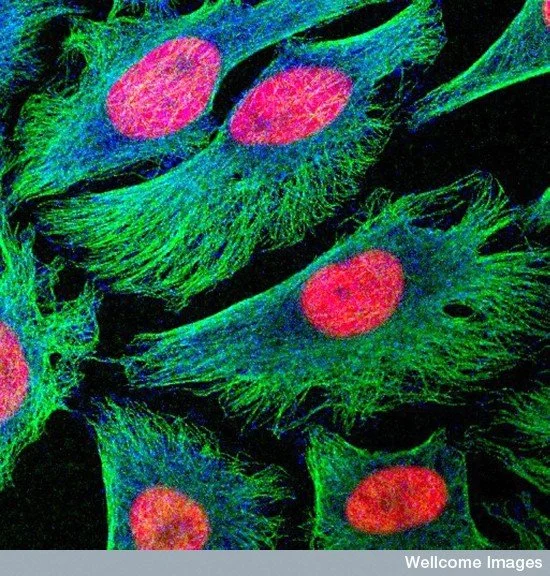
Human HeLa cells in culture. The cytoskeleton is in green. Image credit: Matthew Daniels. Source: Wellcome Images.